

3800 Reservoir Rd, 1st Floor Gorman
Washington, DC 20007
6862 Elm St, Suite 800B
McLean, VA 22101


3800 Reservoir Rd, 1st Floor Gorman
Washington, DC 20007
6862 Elm St, Suite 800B
McLean, VA 22101
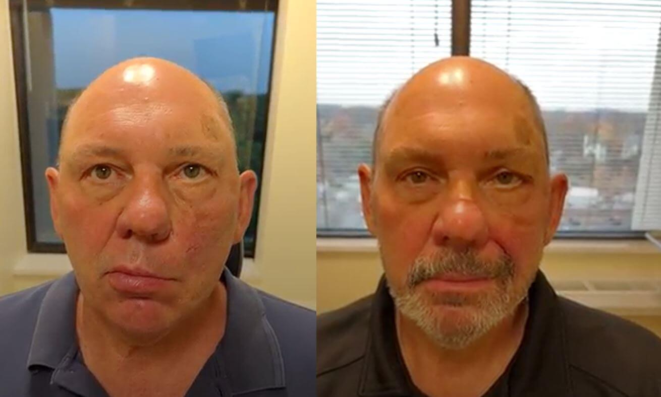
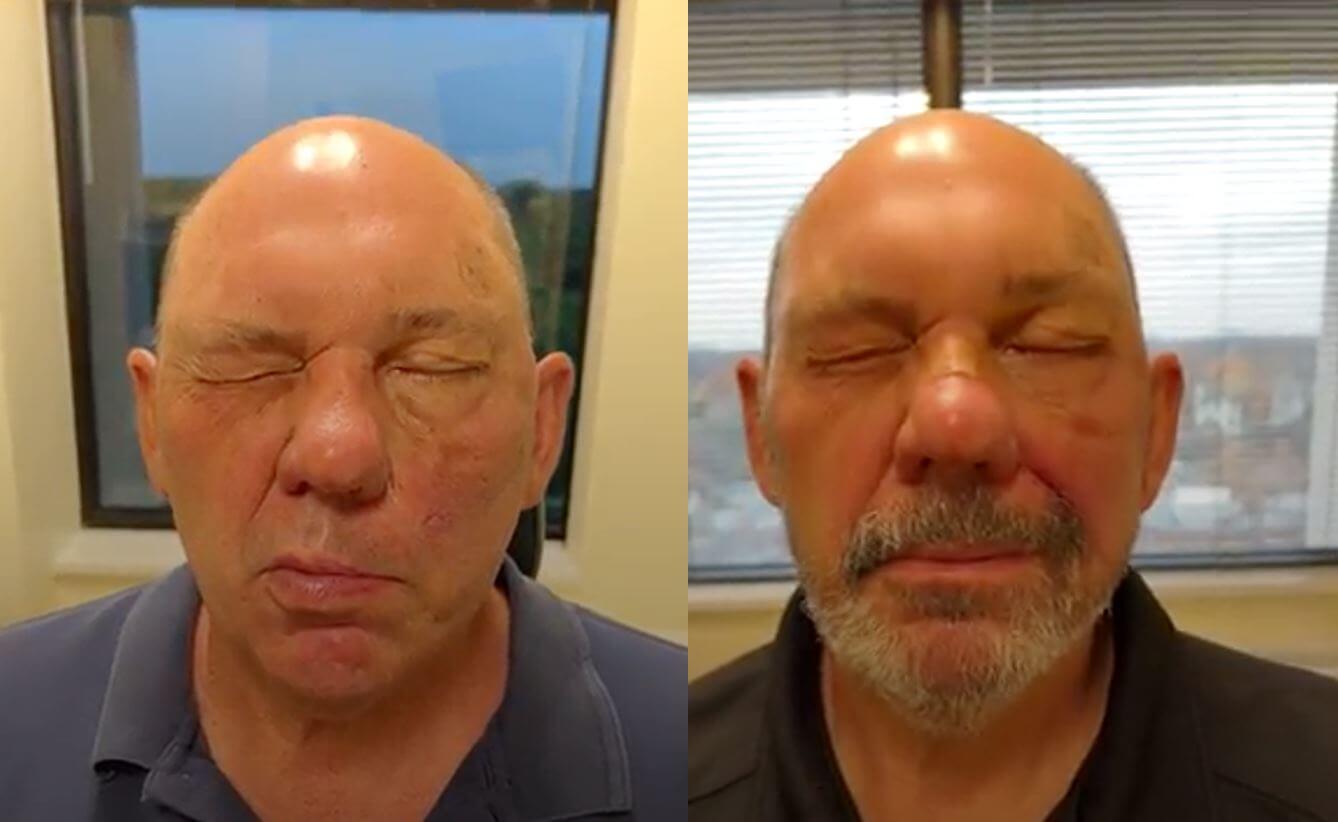
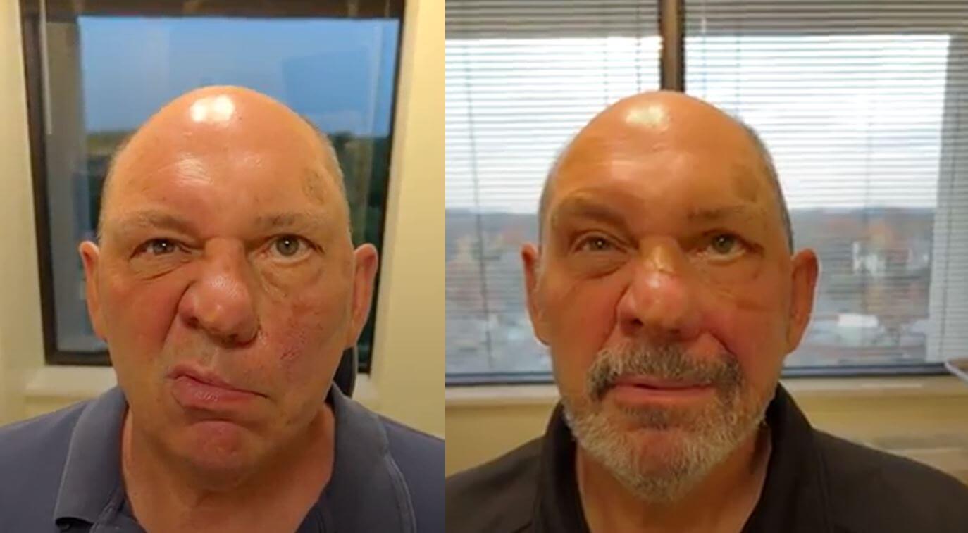
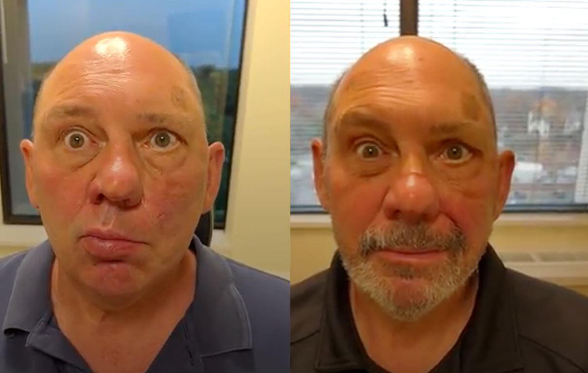
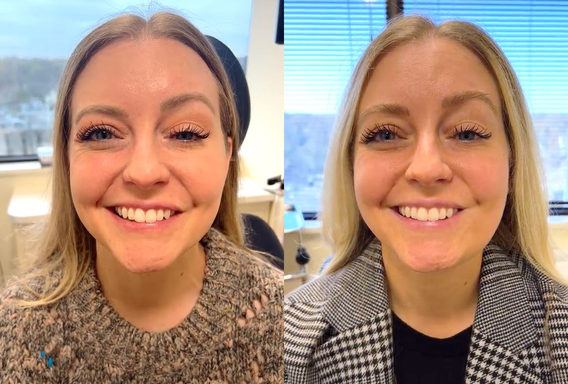
Facial reanimation is the process of surgically correcting facial paralysis; congenital or acquired through trauma or disease. In patients with long-term facial paralysis (more than 1-2 years), the native muscles in the face unfortunately irreversibly lose their function. Therefore, a new muscle must be used in order to restore smile and lower face movement. The purpose of these procedures is to achieve the best functional and aesthetic outcome possible. It may require more than one procedure to optimize the outcome, but the vast majority of patients are able to achieve the goal of improved symmetry and improved smile.
Facial reanimation is a surgical procedure that is performed to correct facial paralysis, whether congenital in nature or acquired through trauma or disease. Facial Botox injections can help retrain the muscles of your face but is only a temporary fix. In patients who suffer from facial paralysis for one-to-two years, the facial muscle function is irreversibly lost. In facial reanimation surgery, Dr. Reilly performs various techniques to achieve the vest functional and aesthetic outcome possible. More than one procedure may be needed to optimize the outcome, but the vast majority of patients are able to achieve the goal of improved symmetry and improved smile. Dr. Reilly is a founding board member of The Foundation for Facial Recovery, a non-profit organization dedicated to improving the lives of patients living with facial paralysis.
Dr. Reilly performs various procedures that can be used for facial reanimation surgery, depending on the specific needs and circumstances for the individual. The options include static tissue repositioning, temporalis tendon transfer, nerve grafting, and partial neurectomy. The incision lines are closed very precisely with small sutures and are well-hidden in the natural folds of your facial anatomy.
We recommend taking a full week off of work or school to adequately rest and recuperate from your procedure. We will see you in the clinic one week after your facial paralysis surgery to ensure everything is healing properly and to remove any sutures. Depending on your occupation, you may be feeling up to returning to work after that first week. You should expect to be taking it easy for the first two weeks after surgery; we recommend no heavy lifting or strenuous exercise for a full two weeks. Avoid any rigorous exercise or contact sports for six full weeks after any facial reanimation operation.
The facial reanimation surgery cost is typically submitted to your insurance company and covered under your medical insurance benefits. We have a team who will help navigate you through the scheduling and authorization process.
There are various procedures that can be used for facial reanimation surgery, depending on the specific needs and circumstances of an individual patient. These may include:
Static Tissue repositioning — This category of procedures involves suspending the corner of the mouth and the fold between the nose and the mouth (laugh line) to an anchor point on the bone just below and behind the eye. This is typically done with a combination of sutures and either the patient’s own connective tissue (fascia lata) or a piece of cadaveric skin (Alloderm), which is used to bridge the gap between the mouth and the anchor point. These procedures are utilized to create better facial symmetry and to address side-effects of facial paralysis including drooling and biting of the lip. The procedure is usually performed through a combination of a small incision in the laugh line and a small facelift incision. The graft is then placed under the skin where it is not visible. Endoscopic equipment is utilized to minimize the incisions.
Alloderm is freeze-dried, acellular human dermis which has been used in reconstruction all over the human body for decades with excellent results. The advantage of Alloderm is that a second surgical site is not necessary for harvesting of the material. Alloderm can be customized just like Tensor Fascia Lata and is readily available. Alloderm tends to integrate well with surrounding tissue and has a very limited risk of rejection.
Tensor Fascia Lata is very strong connective tissue that can harvested from the thigh through a small incision without having any negative functional effects. The advantage of Tensor Fascia Lata is that the material is taken from your own body and further lowers the risk of infection and/or rejection.
Muscle transfers – These procedures involve moving tendons and muscles from one part of your body, usually your legs or abdomen to your face. Some of the tendons that are moved include the temporalis tendon transfer and the gracilis muscle transfer.
∙ The depressor anguli oris (DAO): This muscle attaches to the corner of the mouth and the bottom of the mandible (lower jaw), just to the side of the chin. It is one of the muscles which helps make a frown/express disdain.
Patients with chronic facial palsy from recovered Bell’s Palsy, Ramsay Hunt syndrome, Lyme disease, acoustic neuroma surgery, or trauma, often have too much activity in this muscle on the affected side of their face. This hyperactivity not only pulls down on and restricts the smile, but also can cause tightness, spasm and pain in the cheek and mouth area.
To remedy this downward pull and smile restriction, we have two options:
1.) Inject Botox into the DAO muscle, which helps relax the downward pull temporarily (lasts about 3-4 months)
2.) Surgically resect the DAO muscle, to permanently relax the downward pull and unlock the smile.
Once you are anesthetized, an incision is made inside the lower lip, and fibers of the muscle are identified and removed. We then place a few dissolving stitches to close the incision, and you’ll be given a few days of an antibiotic, as well as a small prescription for pain medicine. Most patients find that they do just fine on extra strength Tylenol, but we do send you home with a prescription for something a little stronger just in case. You can eat and drink normally after the numbing medication wears off (about 2-3 hours) but avoid excessively hot or spicy foods for 72 hours. As you heal, you may develop some swelling on the inside of the mouth over the area where the muscle was removed. This phenomenon is completely normal and resolves with time–most patients feel like they have some swelling (on the inside of the mouth—not noticeable to anyone but you) and mild tenderness for a few weeks after the procedure, and then that fullness shrinks down to a small little “ridge” inside the mouth. After three weeks, patients are encouraged to begin a little gentle scar tissue massage to the area to expedite resolution of the swelling.
∙ Gracilis Muscle Transplant (Free Flap): The gracilis muscle is located in the inner aspect of the thigh. A small portion of this muscle – with its blood supply (artery and vein) and nerve – can be transplanted in the face to replace the facial muscles that allow you to smile. Using highly specialized microsurgical techniques, the gracilis muscle’s artery and vein are attached to an artery and vein in the head/neck region. This connection is critical for the muscle to survive in its new environment in the face. The nerve that moves the gracilis muscle (obturator nerve) must then be attached to a new nerve supply in the face, in order to power the muscle to move. Nerve Options to Power the Gracilis Muscle: The three most common options are described below. More than one option may be proposed to you based on your goals of care.
∙ Contralateral/Normal Facial Nerve via Cross-Facial Nerve Graft: This usually requires two surgeries, unless in certain circumstances where two nerve options are used.
First Surgery: A sensory nerve from the lower leg (sural nerve) is removed and attached to a facial nerve branch on the normal side. The other end of this nerve is then tunneled underneath the skin to rest in the paralyzed face. Because this nerve graft crosses the face from the normal side to the paralyzed side, it is called a cross-facial nerve graft. After the first surgery, we typically wait 6 to 9 months for the nerve signal to grow across the nerve graft, from the normal side to the paralyzed side.
Second Surgery: The cross-facial nerve graft is connected to the nerve that moves the gracilis muscle at the time of the gracilis muscle transfer. The main advantage of the cross-facial nerve graft is that it is the only option that allows for a truly spontaneous emotional smile. The disadvantages are that it typically requires two surgeries, results in numbness of the 5th toe and the side of the foot (sural nerve) and may result in a weaker outcome in older individuals.
∙ Temporalis Tendon Transfer: The temporalis muscle is one of the muscles for chewing (mastication). The trigeminal nerve (cranial nerve 5) is responsible for its activity. As a result, this muscle can be used to provide voluntary facial movement. The procedure transfers the temporalis muscle from the jaw bone (mandible) to the corner of the mouth. The patient then learns to move the face by contracting this muscle. This temporalis transfer is successful in rehabilitating facial movement for those who are not candidates for more advanced facial reanimation (such as cross facial nerve grafts). The procedure is performed via small internal and external incisions between the nose and mouth with very good results. With dedicated exercises, patients are able to obtain voluntary control of the movement of the corner of the mouth, replicating a smile, though this movement does require effort and is rarely spontaneous. Patient satisfaction following temporalis tendon transfer has been shown to be relatively high in several research papers. In a study published in Archives of Facial Plastic Surgery, a mean satisfaction score of 8.5 (possible score of 10) in patients who underwent this procedure.
Nerve grafting — These procedures include moving nerves from different parts of the body to the face, allowing you to better control the muscles. This may include connecting one of the following nerves in the area to the muscles that control the smile and other facial expression.
∙ Masseteric Nerve: The masseter muscle is one of many muscles that help you chew. A branch of this nerve can be reconfigured to power the facial musculature or a new muscle that is transferred to replace the deficient facial movement (see gracilis muscle transplant above). The primary advantage is that the masseter nerve is a strong nerve that is easily accessible during surgery. The disadvantage is that this procedure requires teeth clenching in order to smile. This requires practice, but this learned pattern of movement can become relatively effortless in many patients. For two weeks post-operatively, patients are asked to avoid pressure to the affected cheek, exercise, heavy lifting and vigorous brushing of the teeth in that area. A soft diet is usually prescribed for a period of two to three weeks. The recovery of facial resting tone is usually apparent by 6 months after surgery. Older individuals may require longer periods of time to achieve the desired function, however, the end result is usually comparable to younger individuals.
∙ Hypoglossal Nerve: The hypoglossal nerve moves half of the tongue. A portion of this nerve can be reconfigured to power the facial musculature or a new muscle that is transferred to replace the deficient facial movement (see gracilis muscle transplant above). The primary advantages are that the hypoglossal nerve is a very strong nerve that is fairly accessible. The disadvantages are that there is a small risk of tongue weakness (that can result in difficulty speaking and eating) and there is a risk of inadvertent facial twitching when moving the tongue, such as during eating. Like the masseteric nerve above, practice and exercise are required to coordinate tongue movement for smile, which typically becomes less of an effort over time.
∙ Cross-facial Nerve graft: For patients desiring the best option for restoring spontaneous facial movement, a “cross-facial” nerve graft is the treatment. In this procedure, a nerve graft is harvested from the lower leg (sural nerve graft) through a series of 3 or 4 two-centimeter incisions. The nerve is then connected to one of the facial nerve branches on the unaffected side of the face. The grafted nerve is then tunneled under the skin across to the affected/weak side of the face and left in place for 6-9 months while the nerve fibers brow along this scaffold. This process generally proceeds at a millimeter a day or about one inch per month. At this point, the nerve is connected to a gracilis muscle transplant (see above), so that the spontaneous activation of the muscles on the unaffected side of the face will translate into movement on the affected side, without the need for a volitional movement such as jaw clenching (masseteric nerve transfer) or tongue protrusion (hypoglossal nerve transfer).
All procedures involve a certain amount of risk and limitations. Although the risks of the facial reanimation procedures are relatively low, they do exist. Potential complications from surgery include and are not limited to bleeding, infection, pain, scar, difficulty ambulating (rare for this to be a long-term problem), numbness, failure to improve or worsening in facial weakness/symmetry, and flap failure.
The technique involves first accessing the bone and cartilage support of the nose. This is done with a combination of incisions made inside the nose and in the area of skin separating the nostrils. Next, the underlying bone and cartilage is reduced, added to, or changed to create a newly shaped structure that addressed any post-traumatic deformity and/or nasal airway obstruction.
Once this work is complete, the tissues are then redraped over the new frame and the incisions are closed with dissolving sutures inside the nose and removable sutures on the outside. A small plastic splint is applied to the outside of the nose to minimize swelling and to help maintain the new shape while the nose heals. Soft, absorbent material is most often used inside the nose to maintain stability along the dividing wall of the air passages called the septum.
Follow-up care is vital for this procedure to monitor healing. Anything unusual should be reported to your surgeon immediately. It is essential that you keep your follow-up appointments.
Arrange for someone to stay with you for the first 24 hours if you are not admitted to the hospital for overnight observation.
Go to bed and rest, lying on your back, with your head elevated with 2-3 pillows. You should be lying at a 45 degree angle.
You may be up and around and able to go to the bathroom. You will be able to eat a light meal with assistance.
Take medication only as directed.
Moderate pain after surgery is a normal occurrence, which should be relieved by the pain medication prescription you have been given. Most patients require pain medication for less than 7 days after surgery.
A mild to moderate amount of bloody drainage may occur from any drains that are left in place under the skin surfaces. If drains are left in place, they are typically removed with the first dressing change the morning after surgery.
Gently place ice packs or a bag of frozen vegetables on and around the face for the first 24 hours after surgery (on for 10 minutes, then off for 10 minutes).
On the day after surgery, any dressings should be removed and then replace if there is any evidence of drainage from teh
incision lines.
You may be up and around as tolerated but expect to tire more quickly than usual.
Keep activity and meals light.
You will come into the office for a post-operative check-up and to have sutures removed.
No alcohol for the first 7 days after surgery, which can increase bruising and swelling.
Your swelling and bruising will gradually fade over this time period, but it may persist for up to 3 weeks.
No strenuous activity or heavy lifting for 2 full weeks after surgery.
1. Rest and good nutrition are important healing factors, especially during the first 6 weeks.
2. Numbness and tingling of the incision areas may occur, which resolves with time.
3. The strength and control over the reconstructed smile continues to improve over several years. Over time, however, the brain has the potential to undergo a re-education process (cerebral adaptation) resulting in the production of a naturally occurring, effortless smile even in cases where nerves other than the facial nerve are utilized.
